
AVMs (Arteriovenous malformations)
AVM stands for Arteriovenous Malformation is a tangle of blood vessels, that grew abnormally, and have a risk of bleeding. In almost all cases, it developed during the development of the child in utero. It basically involves two different kinds of blood vessels that should not be directly connected, actually being connected.
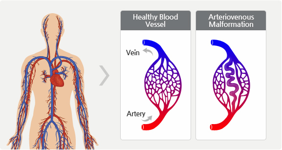
In the human body, there are arteries, which are the vessels that carry blood from the heart to the rest of the body. The arteries are thick-walled and they’re designed to carry high flow and high-pressure blood. Once the blood gets to wherever it’s going in the body, the arteries split again and again until ultimately, they get to the capillaries which are the tiny microscopic vessels where the oxygen is given to all the tissues of the body.
Once the oxygenation has taken place, it’s the veins that take the blood back to the heart from the brain, and the veins are very thin-walled and they’re designed to carry low pressure, slow flow blood back to the heart. So the arteries and the veins are quite different in how they’re built, and they really should not have blood in direct communication.
An AVM consists of an area, where for some reason during development, the arteries and the veins are directly connected by a very tiny network of very small vessels, where there’s basically no pressure drop that’s happening, so you have the high-pressure blood going straight from the arteries into the vein, as a kind of short circuit.
That pressure builds up to the point where there’s a weak spot that eventually ruptures and causes a bleed. Thankfully, the initial bleed is often contained by the surrounding pressure within the skull. This allows a clot to form, which temporarily stops the flow of blood, stabilizing the patient. We still need to treat the AVM before that weak spot ruptures again because every bleed in the brain is a potentially severe risk to the patient.
If the AVM occurs at a very young age and is a very high flow, it can cause other trouble, such as heart failure, etc.
Symptoms of AVM


Symptoms of AVM depend on where it’s located. However, the symptoms after bleeding occurs are:
- Progressive loss of neurological function
- Headaches
- Nausea and vomiting
- Seizures
- Loss of consciousness
Other signs and symptoms of AVM are:
- Weak muscles
- Paralysis in one part of the body
- Problems performing tasks that require planning
- Weakness in the lower extremities
- Back pain
- Dizziness
Diagnosis
Several tools and techniques are there with the help of which we can diagnosis AVM, these are:

Ultrasound
We can determine the pattern of blood flow through the AVM

CT scan or MRI
We can see the AVM’s size and how close it is to normal body parts
an angiogram, to map the AVM’s blood vessels, which will help doctors plan how to block blood flow to it

MRA, or MRA Angiogram
In MRA, we use special MRI techniques to map the AVM without using X-rays. In some cases, a CT angiogram can help them diagnose the AVM.

Standard Angiogram
It uses live X-rays to map the AVM’s blood vessels. This can help doctors plan how to decrease or block blood flow through the AVM.
Treatment Option for AVMs
There are basically three ways to treat an AVM, which are given below:
- Surgery
- Radiation
- Embolization
Surgery
Surgical resection is performed to remove the tangled vessels by cutting them out. The surgeon uses a procedure called a craniotomy to reach the Once the surgeon has access to the AVM, the abnormal arteries and veins are removed. This prevents the AVM from leaking or bursting by redirecting blood flow to regular vessels.
Radiation
When we treat AVM with radiosurgery, beams of highly energized photons (light particles) are directed at the AVM using a tool called a Gamma Knife.
Embolization
In embolization, surgeons insert a type of glue into the AVM through a very thin tube called a catheter. This blocks blood flow into the AVM, which may help limit blood loss during surgery and reduce the risk of bleeding if open surgery is not performed right away.


